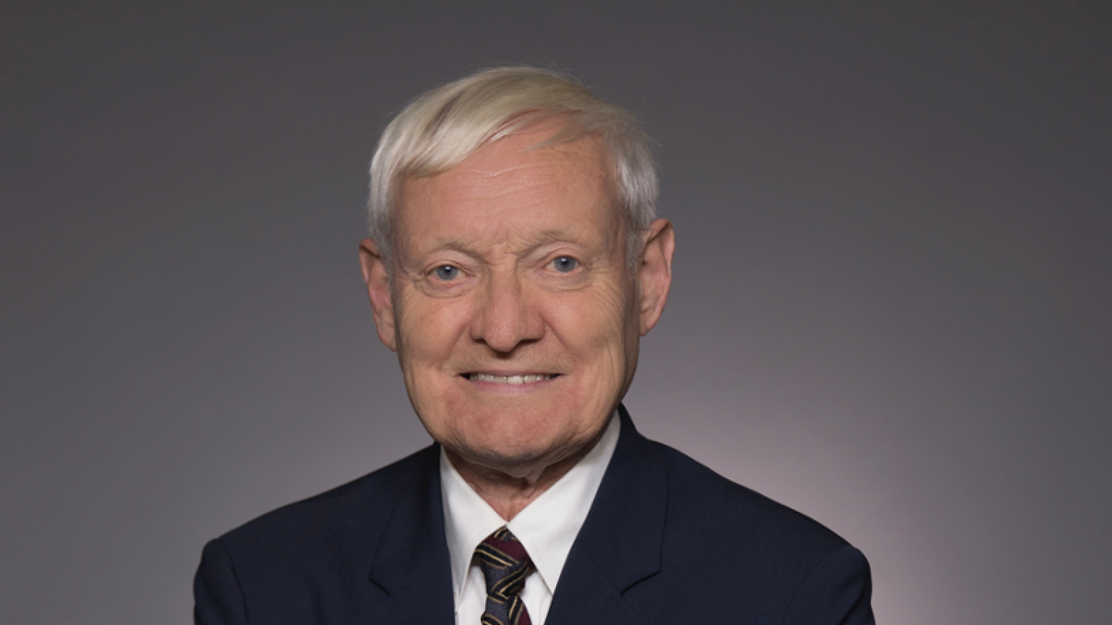
59th Alpheus W. Smith Lecture - Dr. Joachim Frank
The 59th Annual Alpheus W. Smith Lecture, March 22, 2022
Joachim Frank, PhD
Dr. Joachim Frank is a Professor of Biochemistry and Molecular Biophysics, and a Professor of Biological Sciences at Columbia University. He worked on his dissertation under Professor Walter Hoppe’s mentorship at the Max Planck Institute for Protein and Leather Research in Munich (the predecessor of the MPI for Biochemistry in Martinsried) and, in 1970, received his Ph.D. from the Technical University of Munich. A two-year Harkness Fellowship allowed him to visit three labs in the USA for postdoctoral research. After a brief return to Martinsried he spent three years at the Cavendish Laboratory in Cambridge working as a Senior Research Assistant in the lab of Dr. Vernon Ellis Cosslett. In 1975 he joined the Wadsworth Center in Albany as a Senior Research Scientist. In 1985 he joined the Biomedical Sciences faculty in the newly founded School of Public Health of SUNY Albany. In 2008 he moved to New York to assume his current positions. From 1998 to 2017 Dr. Frank was supported as a Howard Hughes Medical Institute Investigator.
Dr. Frank’s lab has developed techniques of electron microscopy and single-particle reconstruction of biological macromolecules, specializing in the mathematical and computational approaches. He has applied these techniques of visualization to explore the structure and dynamics of the ribosome during the process of protein synthesis and the structure and gating mechanisms of several ion channels.
Dr. Frank is a member of the National Academy of Sciences and of the American Academy of Microbiology. He is also a fellow of the American Academy of Arts and Sciences and of the American Association for the Advancement of Science. He was honored for his contributions to the development of cryo-EM of biological molecules and the study of protein synthesis with the 2014 Franklin Medal for Life Science. In 2017 he shared the Wiley Prize in Biomedical Sciences with Richard Henderson and Marin van Heel. In the same year he was awarded the Nobel Prize in Chemistry together with Richard Henderson and Jacques Dubochet.
Cryo-EM of Biomolecules -- Static or in Motion
- Webinar begins at 8:00 p.m. and is open to the public.
- Please preregister to receive the Zoom link for this lecture.
Cryogenic electron microscopy (cryo-EM) has transformed the way we can study biological molecules as it enables us to determine structure of molecules freely suspended in solution (as “single particles”), and to gauge the way they change their shape as they interact with one another in the cell. Freezing, by plunging the sample into a cryogen at liquid nitrogen temperature, is necessary to trap the molecules in a fixed position during imaging and to reduce the damage inflicted by the electrons in the process. Following the introduction of novel cameras for detecting single electrons, near-atomic resolution has been achieved for many molecules and molecular machines of biomedical relevance, including ion channels and receptors. Most recently cryo-EM has contributed in major ways to the development of vaccines and cures for COVID-19.
The standard protocol of cryo-EM does not allow visualization of reactions since the time for sample deposition on the grid and blotting is in the range of several seconds. To study the process of a reaction, as it goes through one or more short-lived intermediates, we have developed a method of time-resolved cryo-EM. Here two reactants are mixed in a microfluidic channel, allowed to react for a defined time (between 20 and 1000 milliseconds), and the reaction product sprayed onto the EM grid as the latter is being plunged into the cryogen. With this approach we have been able to visualize short-lived states of the ribosome in different stages of its work cycle making proteins. Applications in the study of many other molecular machines are possible.
Cryo-EM of Biomolecules – Resolution in State Space
- Lecture open to university students and faculty, 3:30-5 p.m.
- A link to the recording for this session will be posted as soon as possible.
Single-particle cryo-EM offers the opportunity to collect large datasets of images depicting a molecule or molecular machine in thermal equilibrium (at the time immediately before plunge-freezing). Working with our collaborators Abbas Ourmazd and Peter Schwander at U. of Wisconsin at Milwaukee, we have developed a set of geometric machine learning programs, ManifoldEM, capable of mapping the conformational spectrum of the molecule ensemble imaged by cryo-EM, and from this determining the free energy landscape of the molecule. Promises and limitations of this approach will be discussed along with applications.

