
Hammel Group Research
Ultrasensitive Nano-MRI Using Nitrogen-Vacancy Centers in Diamond
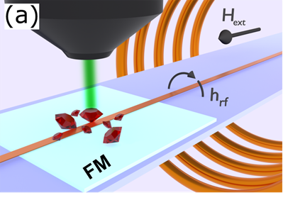
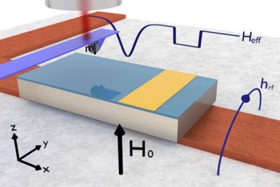
Nitrogen vacancy (NV) centers are fluorescent defects in diamond which are a leading candidate for nanoscale magnetic resonance imaging (MRI) in both biological samples and electronic devices. Their spin-state dependent fluorescence intensity allows sensitive optical detection of magnetic resonance (ODMR) of the NV’s spin 1 system. This unique magneto-optical property enables the measurement of local magnetic fields experienced by the NV center with exceptional sensitivity by measuring changes to the resonance due to these fields. This allows sensitivity to magnetic fields from nearby resonating spins. The atomic size of NV centers promises magnetic field and target spin imaging with unprecedented resolution. Our current work focuses on using NV centers to optically detect the ferromagnetic resonance of proximal ferromagnets with a focus on understanding magnetization dynamics at the nanoscale, and on scanned probe NV imaging of novel magnetic structures including domain walls and skyrmions in chiral magnetic systems. A goal moving forward is to develop protocols for ultrasensitive detection and imaging of high-frequency systems like antiferromagnets, leveraging the unique sensitivity and spatial resolution of the NV system.
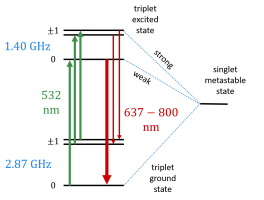
NV: The relevant physics of the spin-1 NV- center is that an incoming 532nm photon causes a spin-conserving transition from the NV ground spin state to the corresponding excited state of the same spin. The NV can then emit a red (637-800 nm) photon upon dropping from the excited state to the ground state. However, the excited ms = ±1 spin state has a higher probability of losing energy via a non-radiative (“dark”) transition through a metastable state than does the excited ms = 0 spin state. The crucial fact that this transition is both dark and not spin-conserving allows continuous illumination under a 532nm laser to polarize the NV center into the “bright” ms = 0 spin state exponentially quickly. Combining this with the knowledge that microwaves (MW) resonant with the NV center cause transitions between the ground ms = 0 spin state and the ground ms=±1 spin states, then the NV EPR can be observed as a reduction in overall PL relative to the PL when off of resonance as the microwave frequency is swept through resonance. This process is known as Optically Detected Magnetoresistance (ODMR). Finally, since magnetic fields along the NV axis cause splitting of the ms = ±1 spin states, then this component of the field can be measured as the splitting of the ODMR peaks in frequency space, and the projection of the magnetic field along the NV axis can then be calculated.
There are several methods to measure the magnetic field depending upon area of interest of any experiment. There are few techniques that we are employing and developing in the lab are briefly discussed below.
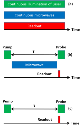
Continuous Wave NV Magnetometry: The word continuous stands for the continuous illumination of laser. In this method, the MW driving and the optical polarization and readout occurs simultaneously. The continuous laser excitation polarizes the NV centers into ground ms = 0 state and the MWs near the resonant frequency drives the NV populations into the less fluorescent ms = ±1 state which cause reduce in PL contrast. A change in local magnetic field shifts the resonant frequency, thereby calculate the corresponding magnetic field. As the laser and MW are turned on during the readout process, the overall sensitivity gives the price for that. In order to further increase the sensitivity, alternatively, the MW frequency can be modulate and measure it using a lock-in technique.
Time-Resolved NV Magnetometry: Time resolved NV magnetometry often known as pulsed NV magnetometry. In order to avoid the optical and MW broadening, this measurement is pursued under some pulsed scheme. In the pulsed ODMR method, a laser pulse is illuminated to NV- to polarize it at ms = 0 state. Then a π pulse is applied for a varying interrogation time τπ (τπ = π/ΩR , where ΩR is the Rabi frequency) and followed by another laser pulse for an optical readout. A change in the magnetic field modifies the spin resonance with respect to the MW frequency, resulting in an incomplete π-pulse and a change in the population transferred to the ms = ±1 state prior to optical readout.
T1 Relaxometry : The NV spins naturally relax at a time scale known as T1 (Spin-Lattice relaxation). One can measure the relaxation time using specific pulse scheme. For a particular NV sample, the relaxation rate mainly depends on the magnetic noise that the NV is experiencing. If the magnetic noise is large the relaxation rate will be higher and correspondingly the T1 will be less. This technique can be used as a magnetic field sensor where the stray magnetic field of the sample is weak.
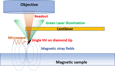
Scanning NV Microscopy: Scanning NV microscopy (SNV) is the technique by which a AFM cantilever designed to contain a single NV center is scanned above a sample. Similar to ordinary magnetic force microscopy (MFM), the tip of the cantilever is scanned in the stray magnetic field in order to build an image of the magnetic texture. However, here the magnetic field at each location is read out optically using the NV center at the tip of the cantilever.
Using this method, an image of the total magnetic field vector as a function of position above the sample can in principle be built up by taking successive scans of the sample with the NV center at three linearly independent orientations. Applications in SNV microscopy include imaging magnetic textures in REIG/GGG systems, particularly measuring current-induced DW/skyrmion motion in such systems.
Ferromagnetic Resonance Force Microscopy
Scanned probe FMR, or FMR force microscopy (FMRFM), is a technique based on magnetic resonance force microscopy (MRFM) in which magnetic resonance is sensitively detected through the magnetic dipole force exerted on a cantilever by means of a micromagnetic tip. FMRFM has demonstrated the sensitivity necessary to study nanoscale ferromagnetic structures. Local properties in thin ferromagnetic films are difficult to probe due to strong exchange interaction causing the lowest energy excitations to reflect global properties in the film - the so-called uniform mode. In contrast, we have used the strong dipolar field from a high-coercivity micromagnetic tip to localize discrete modes directly beneath the tip. By scanning the tip, we have demonstrated real space imaging of the effective internal fields and Gilbert damping using the spatial variation of localized modes resonance. At present, we are actively using this technique to study interfacial spin interactions between ferromagnetic thin film and two-dimensional materials.
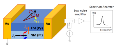
Spin-Torque: Spin-torques generated by spin currents imposed on a ferromagnet can reduce damping, which can be used for magnetic switching or frequency-tunable magnetic oscillators in the GHz frequency range. Spin currents generated by the spin Hall effect offer a simple planar geometry that is easily fabricated and avoids scaling issues that encumber Magnetic Tunnel Junction based structures. Spin-Hall driven auto-oscillations in an extended ferromagnetic film with uniformly applied spin-torque is hampered by nonlinear magnon scattering processes. In order to overcome this issue, spin-Hall oscillators have utilized mode confinement to date by applying electrical current to local nanoscale regions, structural constriction such as nanowires, and local dipolar fields from a micromagnetic particle. Currently, we are using this technique to study the nonlinear spin wave dynamics and damping as well as to engineer the tunable magnon spectrum and spin order control for spintronics applications.
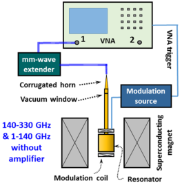
High frequency FMR: FMR has been a major characterization technique for understanding the magnetic excitations and spin dynamics of magnetic materials, but the typical frequency used has been limited below 20 GHz since more specialized techniques are required to process the higher frequencies. Currently, a high frequency (10 - 330 GHz) variable temperature (4 – 300 K) magnetic resonance spectrometer system is being developed in the NanoSystems Laboratory. This instrument can be used for the research in high speed, high efficiency dynamic spin transport driven by magnetic excitations at hundreds of GHz, which includes novel regimes of spintronics utilizing high speed nonlinear spin transport in high frequencies, antiferromagnetic spintronics, and extraordinary dynamics spin conversion in spin-textured materials such as topological insulators and skyrmions.
Cavity FMR: High sensitivity magnetic resonance can be measured using a Bruker EMX X-Band electron paramagnetic resonance spectrometer at a cavity resonance of 9.664GHz. With a compatible cryostat, low temperature magnetic resonance at liquid Nitrogen or liquid Helium temperature can also be measured.
Broadband FMR at variable temperature: We have the ability to measure FMR using customized and commercial co-planar waveguides in a wide range of parameter space: RF frequency from 100MHz to 40GHz; external field as high as 14T; temperature from 4K to room temperature.